Murietta Equine Blog
Lameness Pain

Why Lameness (pain) due to Stifle Joint pathology can be difficult to diagnose.
Abnormal bone development can occur before maturity and not present itself as lameness until early training or mid-career. The size of the bone (cartilage) defect, discipline and training methods can influence when and to what degree the lameness occurs. Some individuals adapt and no lameness occurs.
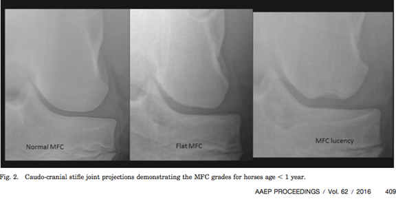
Often with early stifle lameness the radiographs are normal as well as scintigraphy (bone scan) because the early lesion primarily involves the cartilage.
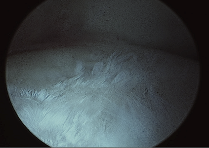
Arthroscopic picture of the end of the femur (medial femoral condyle), illustrating the abnormal cartilage appearing like “floating white grass” instead of its normal smooth glass like appearance. This horses stifle radiographs were normal.
Consider The Following:
- Stifle lameness does not always cause joint swelling.
- Early in the disease process the lameness is often only recognized as a poor performance during high speed maneuvers and can be confused with proximal suspensory, lower back and or SI pain.
- No response of the primary complaint after routine hock maintenance.
- Commonly but not always, stifle lameness is more vivid when the involved limb is on the outside of the circle.
Diagnostics:
- After the lameness from the hock and below has been eliminated as the source of pain, even if the stifle radiographs are within normal, block the stifle.
- When blocking the stifle joints (3), an ultrasound -guided technique is preferred for consistent accuracy. It can take up to an hour before the block can change the lameness.
Prognosis For Stifle Lesions:
- The prognosis improves with early detection.
- The accuracy of the diagnosis dictates the treatment and prognosis.
- Intraarticular corticosteroids and continued exercise often decrease the prognosis.


Comments